How Metabolism Can Shape Cells’ Destinies
Introduction
Each of us starts life as a single cell. To develop into a complex, multicellular being, that cell must divide, and then those cells must divide again, and again — and then these stem cells start to specialize into different types, with different destinies in our bodies. In the first week, our cells reach their first turning point: They must become either placenta or embryo. Then, in the developing embryo, cells form three primary layers — ectoderm, mesoderm and endoderm — which, over time, become skin, neurons, heart, gut, and so on.
These determinations of cells’ fates — what type of specialized cell they will become — occur in stages throughout embryonic development. Because each cell type has a characteristic pattern of gene activity, scientists assumed that the decisions cells make are dictated by genes: specifically, networks of genes that turn each other on and off, initiating a cascade that forms the correct types of cells in the correct order.
But genes are not the whole story. New research has shown the extent to which cell metabolism — the chemical reactions within a cell that provide energy and materials for growth — has an important, underappreciated role in directing cell fates.
“Metabolism is more than just housekeeping in stem cells, especially embryonic stem cells,” said Jan Żylicz, a developmental biologist at the University of Copenhagen. “It’s a crucial pathway that regulates decision-making processes.”
In the course of their whirring biochemical activity, cells not only produce energy but also synthesize metabolites: molecular biological building blocks, such as amino acids, nucleotides, carbohydrates and lipids. In the last decade or two, with the development of better methods for measuring metabolites in cells, there has been a surge of interest in the various ways these small molecules regulate gene activity, and in particular cell fate and development. Now, studies suggest that their presence or absence — which can be influenced by external factors, such as environment and diet — can determine the fate of a cell and, in turn, the development of an embryo.

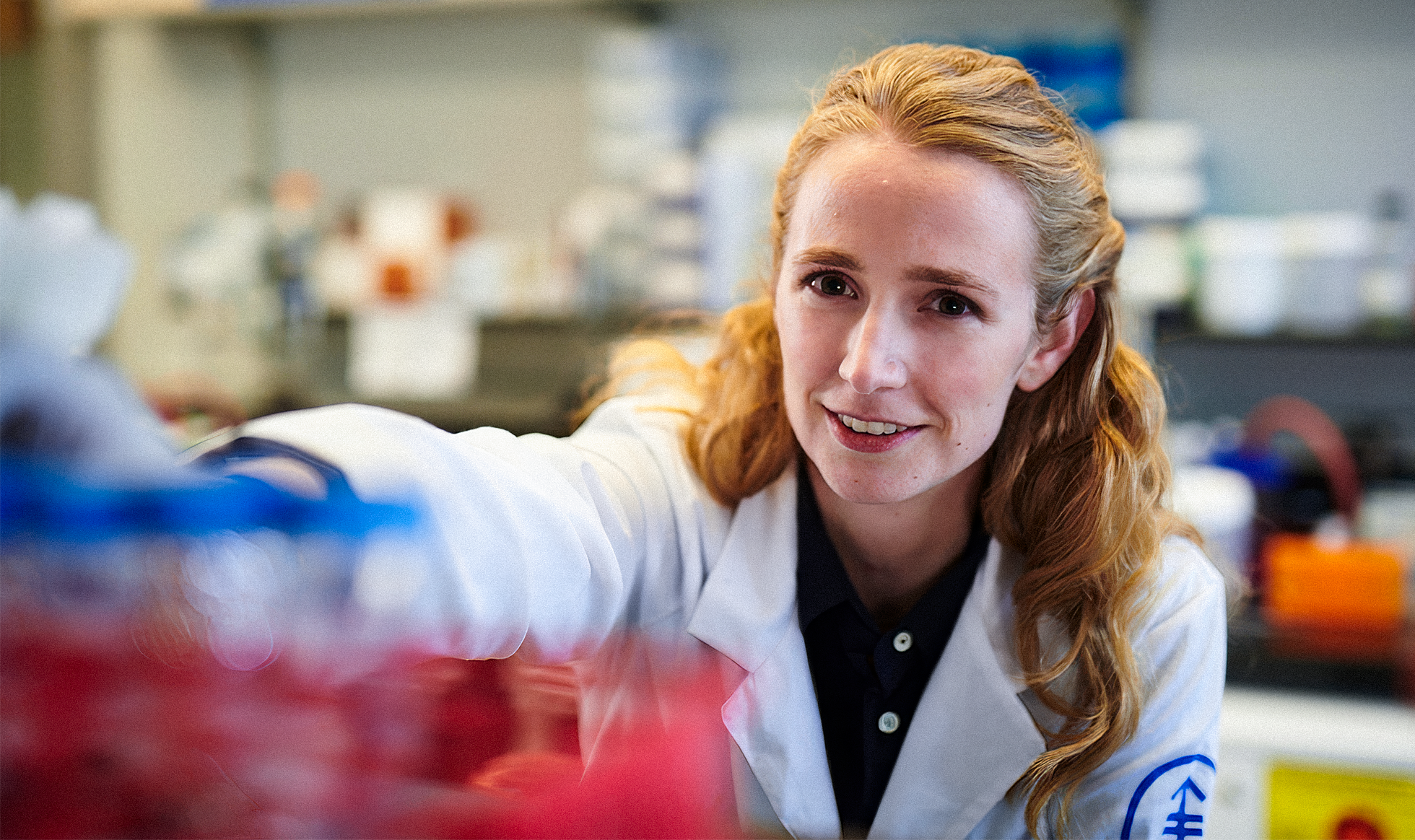
The developmental biologist Jan Żylicz has seen how a single type of metabolite can change the fate of a cell during the earliest stages of human development.
Courtesy of Jan Zylicz/UCPH, Denmark
“Beyond the bioenergetics, these by-products of metabolism are used for regulating specialized programs as well,” such as cell differentiation and the formation of an embryo’s three layers, said Berna Sozen, a developmental biologist at Yale University who recently published research in Nature showing how glucose metabolism influences the earliest stages of embryonic development. “The possibilities are so exciting. It really changes the way we think about developmental biology, the way we think about how our own life starts.”
Scientists have traditionally believed that all the instructions a cell needs to become a particular type are encoded in its DNA. In that case, when a stem cell differentiates, part of that execution involves turning on the genes that encode that cell type’s metabolism, said Jared Rutter, a biochemist at the University of Utah. But studies now show that the operation can run backward: The cell tests whether it has the materials in its environment. If it cannot execute the metabolism, then it won’t become that cell type, in spite of signals to differentiate. “It’s a revolution in my thinking of how metabolism influences things,” Rutter said.
The body of work overturns assumptions about the pure dominance of genes during development and helps us understand the factors that contribute to an embryo’s survival, cell death and even cancer.
“Almost any question is on the table,” said Lydia Finley, a cancer biologist at Memorial Sloan Kettering Cancer Center in New York. “The field of metabolism and development is really developing now, which is super exciting, because it’s early, early days.”
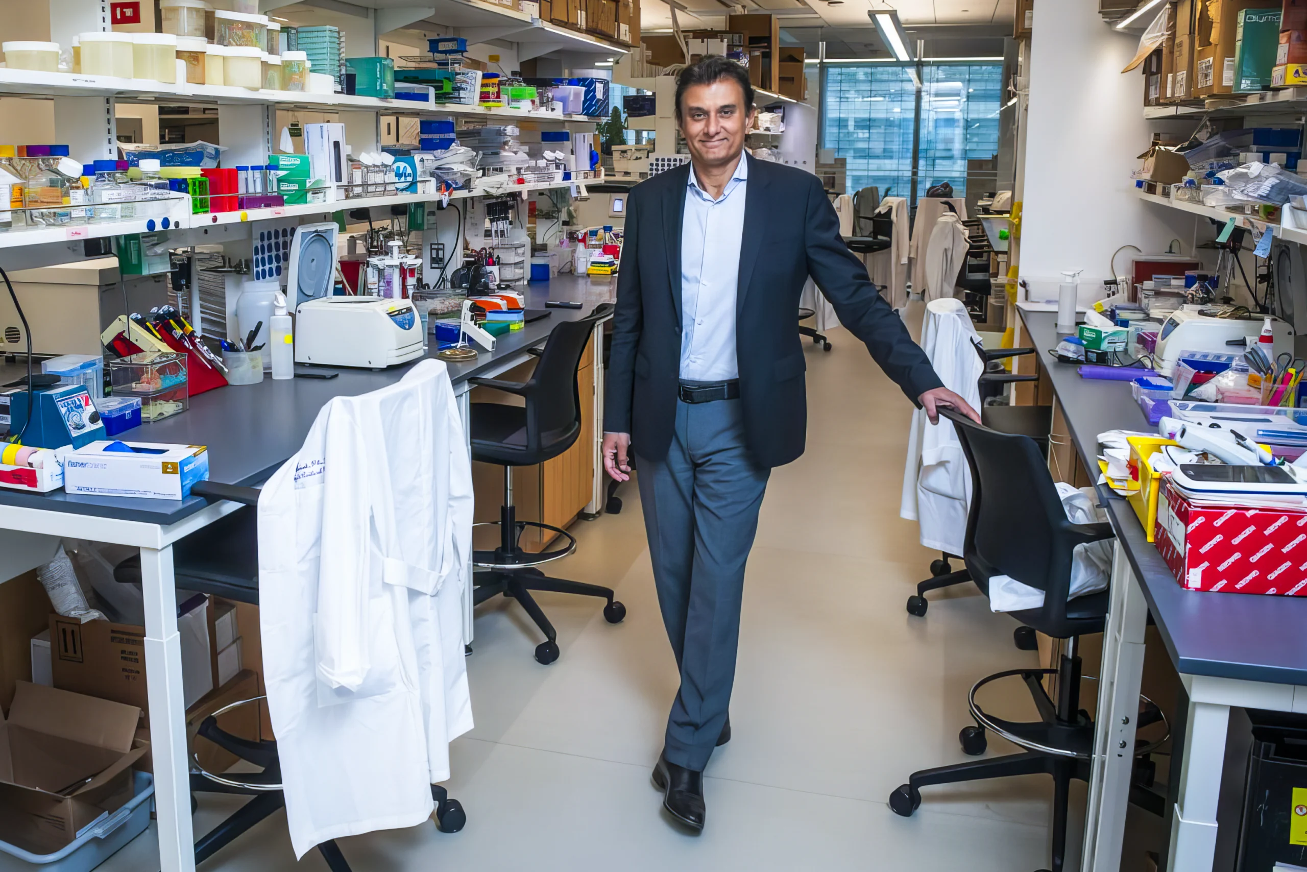
Lydia Finley of Memorial Sloan Kettering Cancer Center discovered that the anti-tumor protein p53 helps prevent cancer by managing a cell’s metabolic state.
Memorial Sloan Kettering Cancer Center
Early Signals
One of the most striking examples of how metabolism can drive cell differentiation comes from a humble slime mold. When Dictyostelium has plenty of nutrients in its environment, it happily grows and divides as a group of single cells. But when food dries up, a change occurs: Individual cells aggregate and form a sort of multicellular slug, which crawls as a single unit and forms fruiting bodies to reproduce. While food availability is the obvious trigger for the change, until recently no one knew how exactly it flips the switch from unicellularity to multicellularity — a form of cell fate — at the molecular level.
Four years ago, the immunologist Erika Pearce and her team studying cell metabolism at Johns Hopkins University discovered how this switch is metabolically driven. Under starvation conditions, Dictyostelium mitochondria generate a burst of reactive oxygen species — small, unstable molecules that can damage proteins and DNA, and can also act as signaling molecules. To protect itself from its own mitochondria, the cell produces an antioxidant called glutathione.
Glutathione doesn’t come out of nowhere: It requires the nutrient sulfur. A starving slime mold cell shunts all of its sulfur into glutathione production. That means there’s no sulfur left to build iron-sulfur complexes, without which the cell can’t make new mitochondria. Therefore, the slime mold “has no choice but to become multicellular,” Pearce said. It can’t grow and spread on its own anymore, so it forms a slug and heads off in search of food.
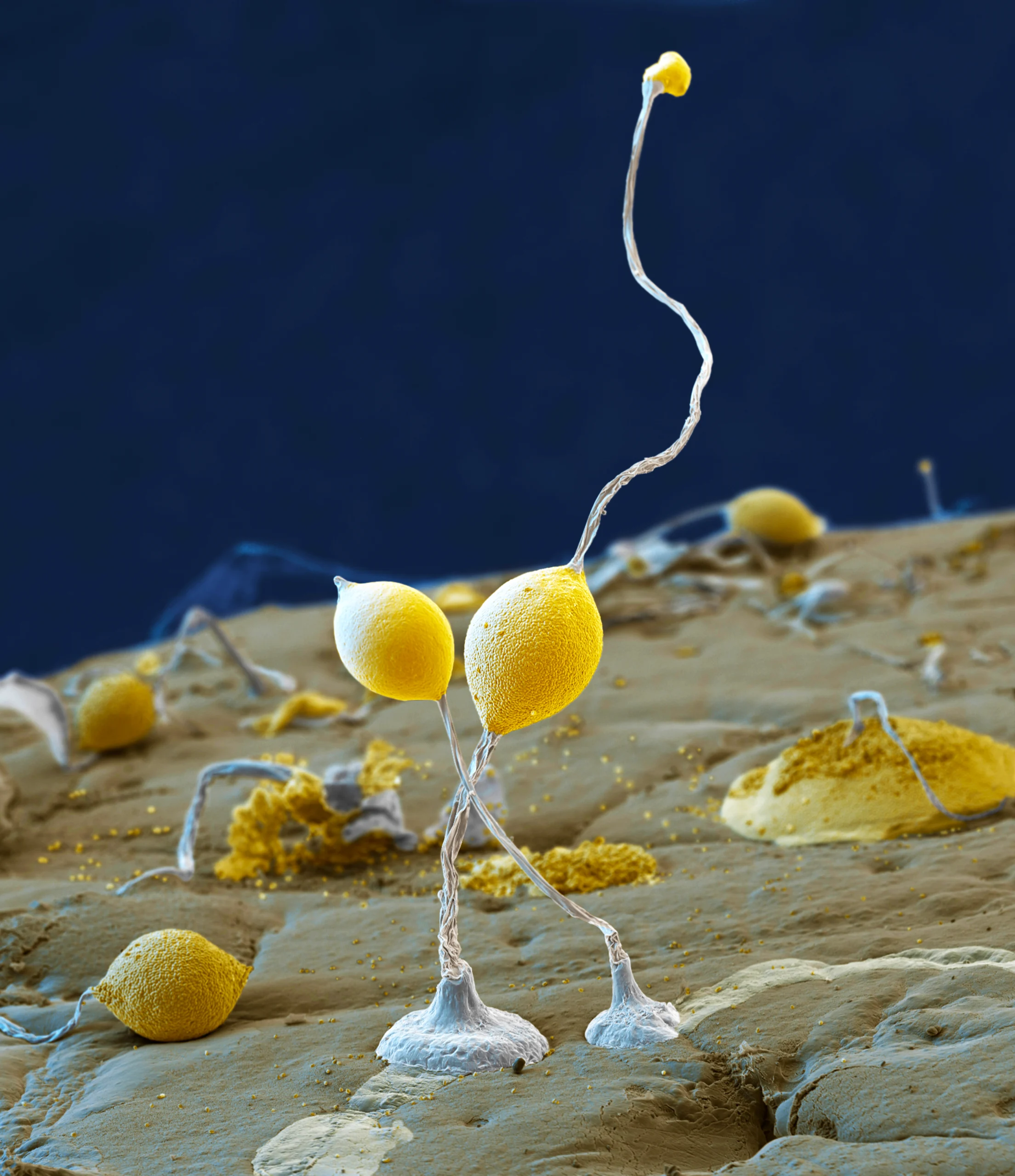
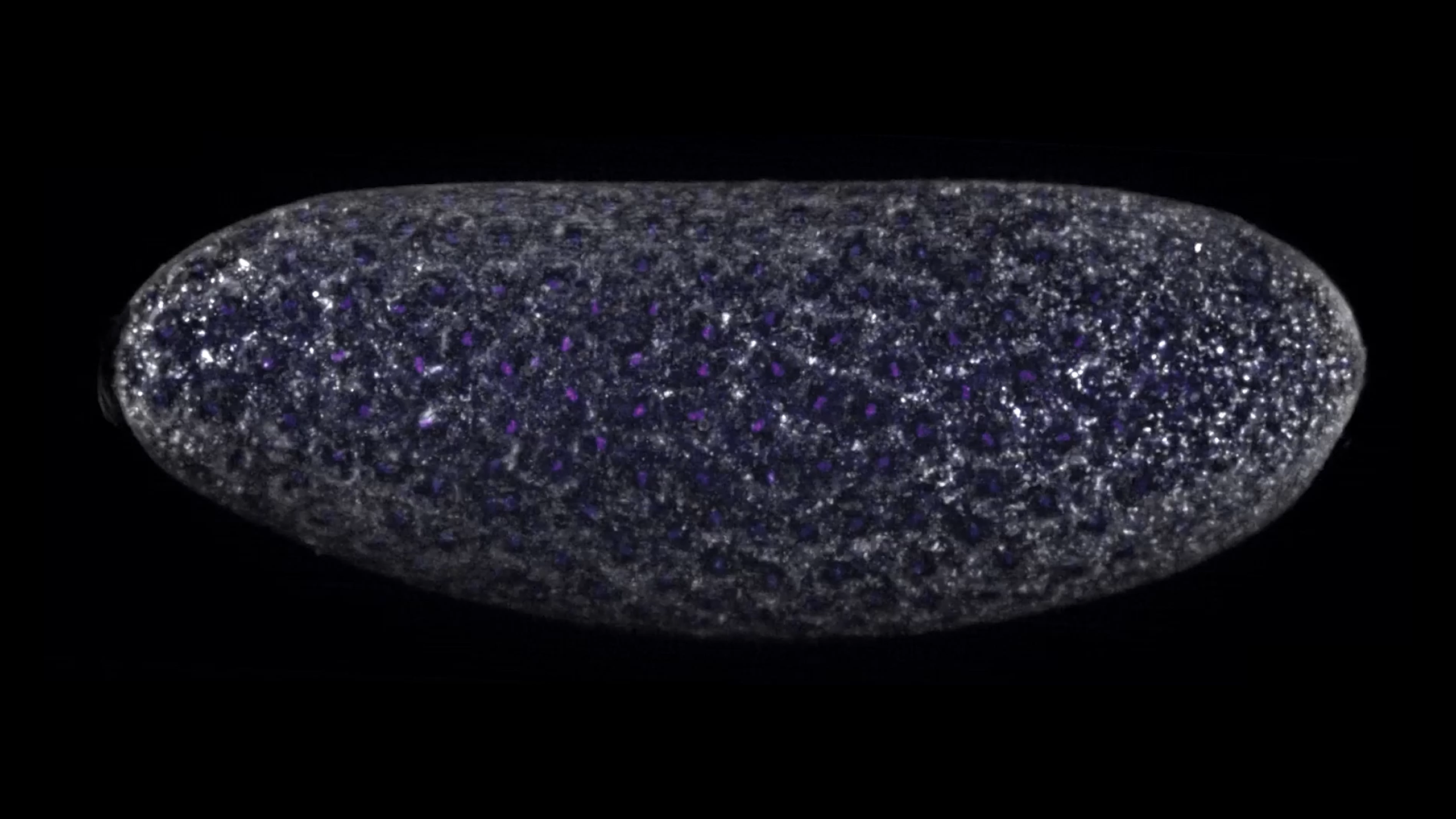
When food is scarce, the Dictyostelium slime mold abandons a unicellular lifestyle and switches into a sluglike multicellular form with fruiting bodies (pictured here). At a cellular level, this change is driven by the lack of a single nutrient: sulfur.
Eye of Science/ Science Source
“Metabolism was driving that entire phenotype, and whether or not you had food there — that is probably still the most fundamental driving force,” Pearce said. “Every single one of our cells is probably subjected to that as well.”
This discovery showed that a cell’s metabolic state can trigger a signaling cascade that completely changes an organism’s form and behavior. However, understanding how cell metabolism is translated into a developmental signal in organisms more complex than slime molds had taken decades of work.
Back in the 1990s, the biologist Navdeep Chandel was a graduate student working on a mitochondrial enzyme called cytochrome c oxidase. “I was a pretty confident young lad thinking I know what cytochrome c oxidase does: It takes an electron from cytochrome c and gives it to oxygen,” he said — a key part of the mitochondria’s process for creating cellular energy in the form of adenosine triphosphate (ATP). But surprisingly, in 1996, researchers discovered that if cytochrome c is released from the mitochondria, it sets off a cascade of signals that triggers cell death — also a kind of cell fate decision.
“So it [cytochrome c] has a second function, a moonlighting function, you could call it,” Chandel said. That was the first hint that mitochondria were doing more than just providing ATP: They were also influencing cell decision-making. Chandel, now a mitochondrial biologist at Northwestern University Feinberg School of Medicine, has been working on elucidating mitochondrial signaling ever since.
Working with human stem cells more than a decade ago, he discovered that mutating a critical mitochondrial enzyme prevented the cells from differentiating into fat cells as they should have. In 2013, his lab showed that reactive oxygen species generated by mitochondria were essential signals in mouse skin development. Then, in 2023, in an experiment published in Nature, he and his team again found that cell specialization could not happen without healthy, functioning mitochondria. In a mouse model, stem cells with defective mitochondria unleashed a stress response — a cascade of molecular signals that activated stress-response genes in the nucleus — and then the cells stalled out, unable to become lung cells. The mice’s lungs failed to develop, and they died.
Navdeep Chandel, a biologist at Northwestern University, has spent his career elucidating how mitochondrial signals affect cell specialization and animal development.
Northwestern Medicine
The stress response, Chandel concluded, was an emergency message to the nucleus to stop development when the mitochondria encountered a metabolic problem.
“When we started these experiments, most people would say, ‘Oh my God, what a dumb experiment, you’re going to get dead cells,’” he said. “But hang on. We haven’t seen that. We’ve seen specific defects — the defects being [cells] not differentiated. I think that’s quite cool.”
Over the past few years, other research projects have independently tied mitochondria’s emergency stress response to cells’ failure to differentiate. For example, in fruit flies, defects in metabolic enzymes in a subset of tissues can trigger a stress response that halts the growth and development of the entire animal. By genetically blocking the stress response, the researchers reversed the effect.
Most recently, in February 2025 in Science, the endocrinologist Scott Soleimanpour at the University of Michigan found that in mice with defective mitochondria, beta cells (special cells that produce insulin) were de-differentiating — losing their identity as beta cells and reverting to a more immature state. By inhibiting the stress response, his team could get the beta cells to re-differentiate, much as Chandel could restore the lung cells in his mice if he suppressed their stress response.
Researchers already knew that mitochondria under stress can send signals to other parts of the cell. These studies help clarify the message. “The animal knows there’s something wrong at a metabolic level, and it releases signals to slow down development,” said the geneticist Jason Tennessen of Indiana University, who led the fruit fly studies.
The research has flipped how Tennessen thinks about the relationship between genetics and metabolism. “Instead of thinking about the gene expression networks just happening to interact with metabolism, it’s really metabolism driving [developmental decision-making],” he said, “and gene expression networks are the tools by which that occurs.”
This idea — that cell metabolism is an integral but unheralded part of the developmental process — isn’t fantastical. In another field of biology, epigenetics, researchers have already detailed the process by which metabolites turn genes on and off. But they needed the work of developmental biologists to connect more of the dots.
The Metabolic Nucleus
Nearly all the different cell types in your body — liver cells, heart cells, skin cells, beta cells, and so on — contain the same genome in their nuclei. What differentiates them is how the gene activity is regulated. In each cell type, a different set of genes is expressed to make the proteins and RNA that allow them to function properly in their respective roles in a mature body.
Epigeneticists who study this process have, over the past few decades, elucidated a complex system by which proteins and enzymes activate or repress certain genes. The meters-long strand of DNA in every cell is wound around proteins called histones. With the help of specific enzymes, molecules that scientists call “chemical modifications” or “epigenetic marks” attach to the histones and cause the DNA to unspool, exposing different genes for activation. These modifications can thereby activate some genes and deactivate others, influencing the biochemical processes in a cell and therefore the functions that cell performs.


Mark Belan/Quanta Magazine
“Those chemical modifications that decorate [histones] and modify gene expression — they’re metabolites, full stop,” said Finley, the cancer biologist. “Chemical modifications themselves are metabolites, and their removal is dependent on metabolites.”
Fifteen years ago, when Kathryn Wellen was a postdoc studying cancer cells, she discovered that the epigenetic marks on histones change in response to the presence of nutrients. When food is plentiful, mitochondria make a metabolite called acetyl-CoA. It diffuses into the nucleus, where the genome resides, through large pores. There, enzymes break down the metabolites into epigenetic marks known as acetyl groups and place them on histones to activate one set of genes. However, when the cells are starving, enzymes strip off the acetyl groups. Some of those acetyl groups are turned back into acetyl-CoA and consumed for energy, while others are recycled to activate a different set of genes.
Clearly there’s a lot of metabolic activity occurring in the nucleus. Wellen wondered whether the nucleus had its own unique metabolism and could therefore be considered a “metabolic compartment.” Working with Nate Snyder, a biochemist at the Lewis Katz School of Medicine at Temple University, Wellen and other researchers developed new methods to measure metabolites in different parts of the cell and saw that metabolic activity in the nucleus is not identical to activity occurring elsewhere.
“Although that may sound obvious, it was not,” Wellen said. The nucleus’s metabolic activity was specific to the functions in that compartment, including epigenetic activity. “There are a lot of metabolic enzymes that are actually in the nucleus and are dynamically regulated in the nucleus,” said Wellen, who now heads a lab at the University of Pennsylvania. “We were really excited to find that.”
This idea of the nucleus as a metabolic compartment was foundational to understanding how metabolism impacts embryonic development. In early embryonic cells, as developmental decisions are made that direct cells to become ectoderm, mesoderm and endoderm, all of the epigenetic marks on the histones get repositioned. They can be removed, added and relocated to activate certain genes and repress others.
“What is intriguing is that all of this is associated with a massive accumulation of metabolic enzymes in the nucleus,” said Żylicz, the developmental biologist. These enzymes make molecules, which then activate other enzymes that remove epigenetic marks and lay down new ones as cells grow, divide and take on different fates.
During this period, the cell moves many enzymes from the cytoplasm and mitochondria to the nucleus. That way, the metabolites necessary for gene activity can be produced locally, in the nucleus, where they are needed, Żylicz said. “The moment where you reprogram the epigenome — that happens to be the same time when you’re also really using this nucleus as a metabolic compartment.”
Early in human development, the embryo is a ball of cells. The cells on the outside form the placenta; the cells on the inside form the embryo. The major difference between these two types of cells is in the activity of metabolic genes. Recently, Żylicz’s team pinpointed differences between these cells in alpha-ketoglutarate, a well-studied metabolite, and showed that the metabolite accelerated the differentiation of stem cells into cells that will become the placenta.
Alpha-ketoglutarate not only controls differentiation in stem cells; it does the same in cancer cells, Finley’s team and other groups found a few years ago. They were studying p53, a protein that is well known for its anticancer effects; its gene is the most commonly mutated gene in human cancer. Their study, published in Nature, found that p53 caused alpha-ketoglutarate to accumulate; this alpha-ketoglutarate altered the fate of the cancer cells so that they were less likely to form tumors. This was striking and unexpected because researchers had assumed that p53 has an anticancer effect by directly regulating the activity of genes. It also works by altering metabolism.
“This is particularly exciting because if changing metabolism can change cell fate in a meaningful way, there is the possibility that you might be able to manipulate that therapeutically, where aberrant decisions of differentiation are causal for the disease — like in many forms of cancer,” said Rutter, who was not involved in the study.
In some ways, this interplay between metabolism and genes is obvious: We know that life is influenced by both its genes and its environment. This new, exciting field of research shows at a molecular level how the materials available to our cells influence their fates, and ours.